1480 Hunt Club Road, Ottawa, Ontario, K1T 1M6
1480 Hunt Club Road, Ottawa, Ontario, K1T 1M6

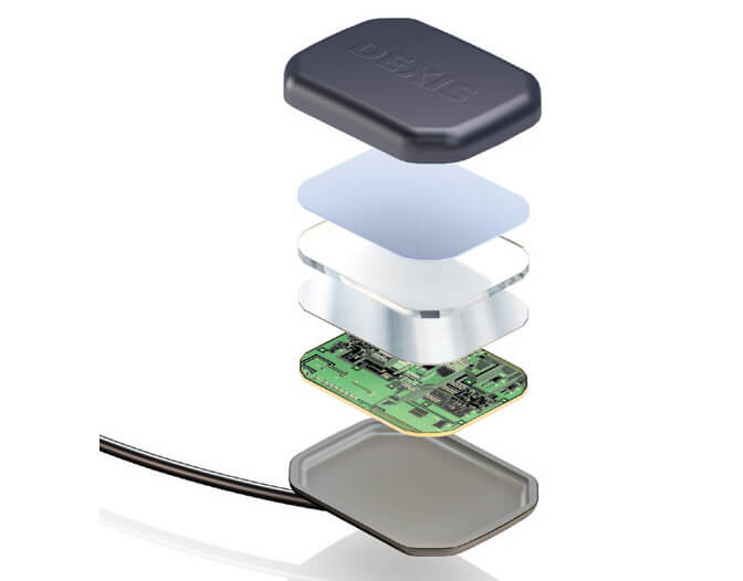
The DEXIS system provides you with all the benefits of direct digital dental radiography. The system is a direct-USB digital x-ray with an optimal performing single CMOS sensor that is directly connected to the computer. This single sensor has the ability to take horizontal and vertical bitewings and every peripheral, removing the discomforts and expensive cost associated with multiple sensors. The DEXIS is different from many direct digital systems because unlike them, it makes use of a direct USB connectivity that is wholly integrated, eliminating the need for a docking station or separate adaptor. As a result of the fact that the connector is gold platted, the USB durability has also increased. There is a wise angle cable exit on the sensor to help patient manage the procedure better.
It makes the cable flexible to increase reliability and reduce stress. There are four beveled corners as well as a round and smooth casing. This makes sure that sharp edges are not found, while the smaller angle dome is essential for accurate posterior placement. Lastly, attached to the inner wall of the rear housing is a protective shield to keep the patient safe from X-ray back scattering. The design of the sensor was made bearing in mind the clinician, technician, and patient.
The DEXIS system can easily be employed in any dental practice. There is an automatic synchronization of the images with the records of the patient, creating an easy access to the patients chart and image.
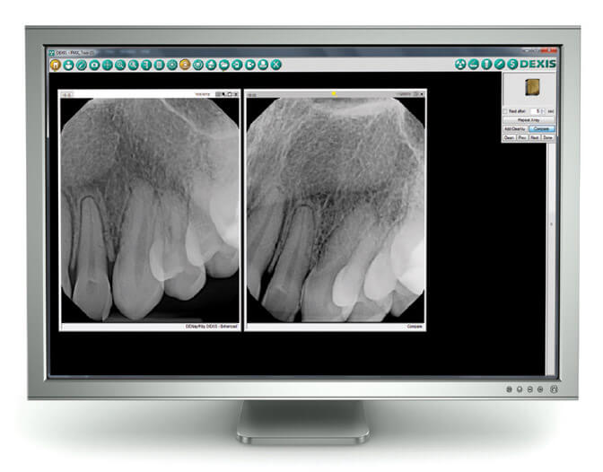
The charting program offers preview thumbnails as well as full-screen images that are clinically relevant.
To view the full-screen images, click on the thumb nail image displayed in the charting program.
Management software which is the DEXIS system’s integrator window can be positioned and sized to fit the screen layout optimally within the practice. It also runs perfectly on the background, without a display of the window. These features make sure the DEXIS system functions well with dental practice software and the work style of the members of the dental team.
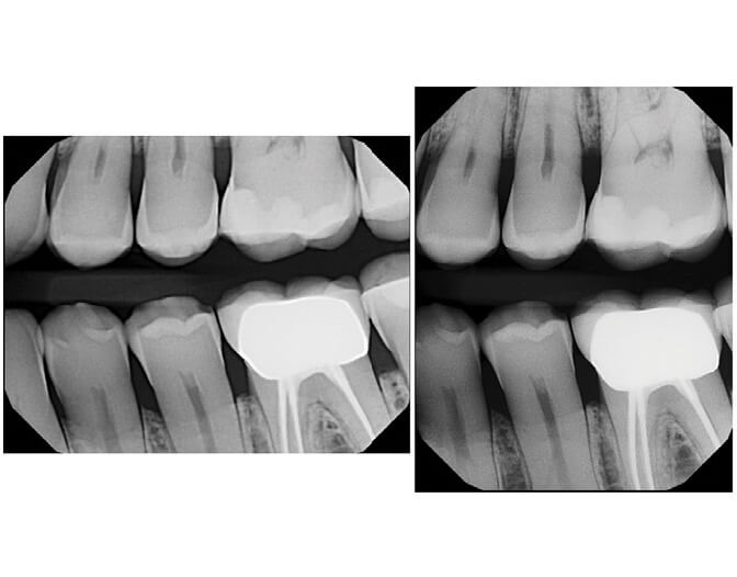
When choosing a digital dental radiography system, the ability to display top quality images is an essential factor. DEXIS employs PureImage technology in forming clear images. The beam from the x-ray is transformed into visible light through a top quality cesium iodide scintillator, which guides the X-ray through its microcolumnar structures. The light is then transmitted to the surface of the CMOS sensor by fiber optics, enabling a high signal-to-noise ratio that brings about pure images with almost no form of visual noise. The active part is maximized at the pixel level by the high resolution CMOS sensor, bringing out even the minutest detail. The improved quantum efficiency of the sensors boosts efficiency and reception when capturing images across various ranges of radiation settings. Delicate variations in densities can be observed through the 14-bit analog-to-digital converter that generates 16,000 shades of grey. The generated images are 2.2 megapixels and can be viewed in very large sizes. The great quality of DEXIS system images can be seen when the images are zoomed in without any loss of pixilation or quality.
The DEXIS system is likewise made to help dental practices function smoothly and with ease. It is very easy to learn and make use of the system. The system is designed in a highly intuitive manner, and most common processes are finished in 1 or two clicks. This feature makes it possible to spend just 5 minutes instead of 25 minutes from the beginning to the end the FMX procedure.
The direct digital CMOS sensor senses radiation and automatically saves the image with date, correct orientation, and tooth numbering. The images are available at once, removing the time hitherto spent on developing and setting up traditional x-rays. This makes it possible for an image to be captured again if there is any issue with the first image. The big images displayed on the screen and other enhancement tools increases the possibility of a more effective communication with the patient and offers support for clinical diagnosis. Due to the centralized image system, there is an easy room for interaction between clinicians through emails and exporting of the images.
The importance of top quality dental radiography images to both the clinicians and the patients can’t be overemphasized as it contributes directly to treatment planning and diagnoses. As a result of its increased clarity, Storing, lower cost, sharing, and ease in manipulating images, Digital dental radiography is presently the most preferable and best option for dental practices. Direct digital systems of high quality like the DEXIS systems, offers an awesome experience for both patients and clinicians.
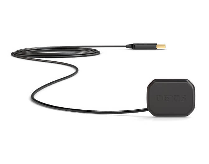
DEXIS Platinum high performance single CMOS sensor connects directly to the computer via USB.
The DEXIS single-sized sensor’s design allow for the capture of vertical and horizontal bitewings.
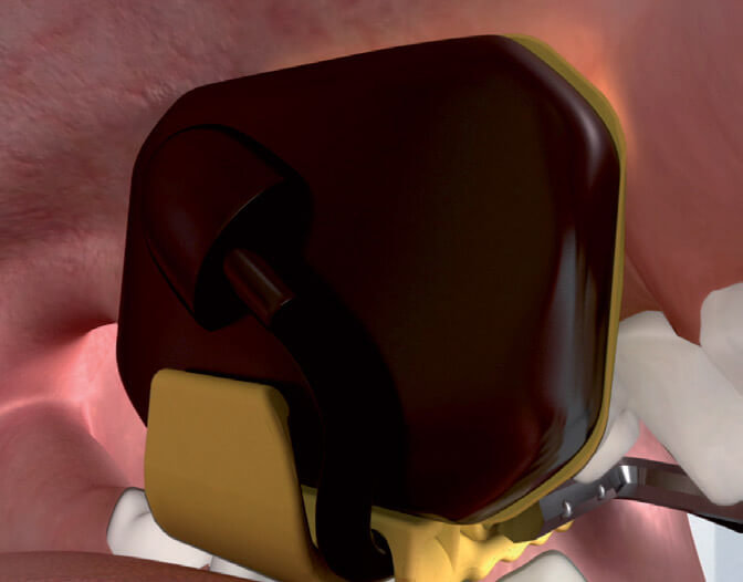
Cable exit placement and smooth, beveled corners help patients better tolerate radiographic procedures.
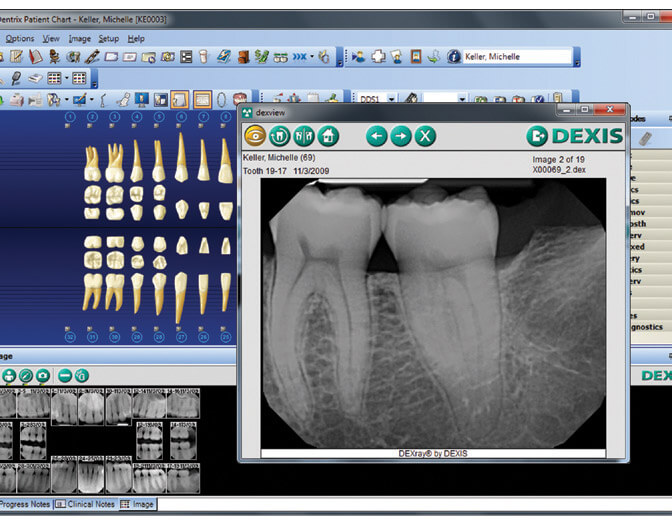
DEXIS Integrator allows for image previews and image enlargement directly from the practice management chart.
PureImage technology delivers clear, highly diagnostic images.
When images are enlarged, there is no loss of quality or pixilation.
*Extra fees may apply.
Reserve by phone or online: